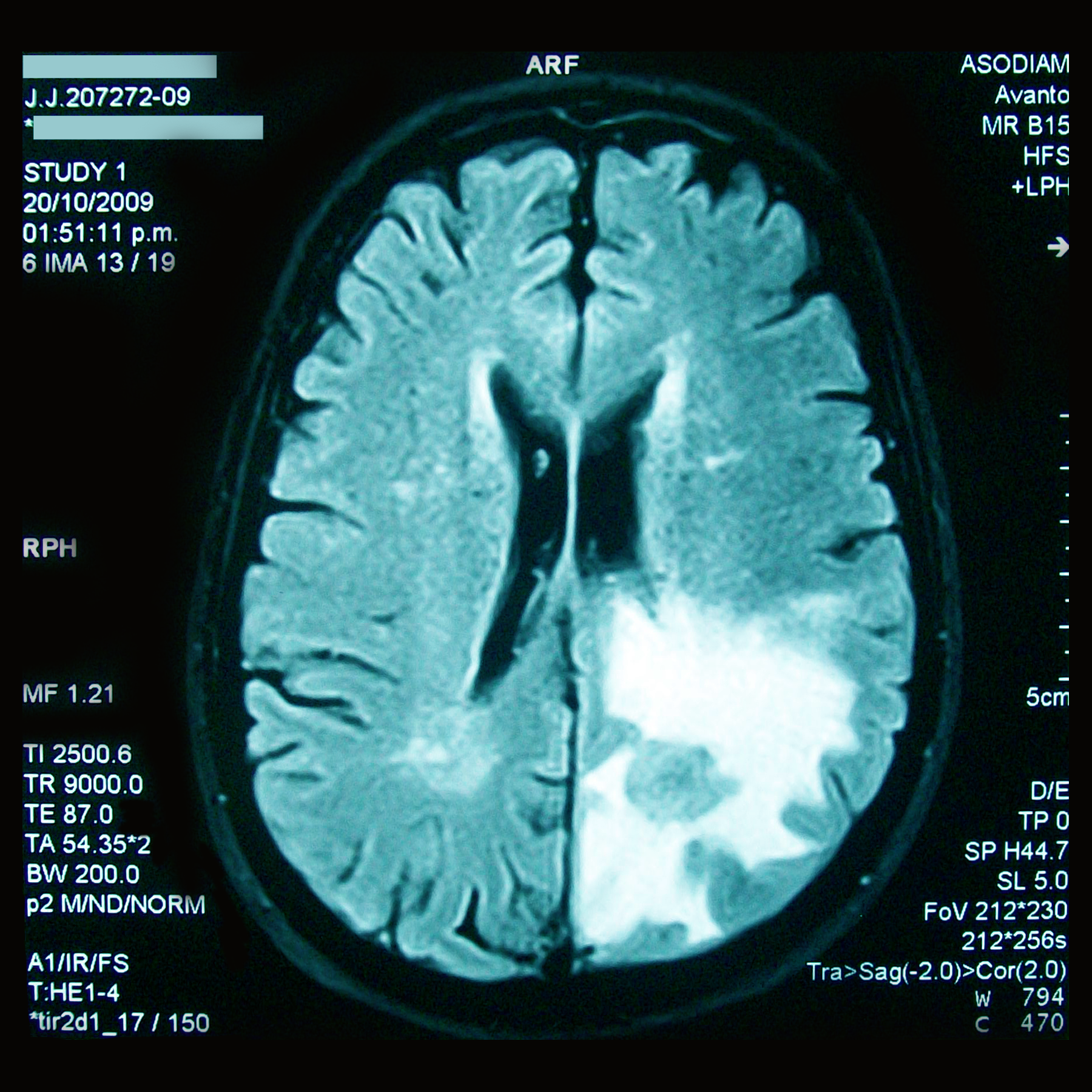
Imaging methods
This edition of the Forum is focused on imaging methods applicable to problems in physiology and biology. The aim is to provide new points of contact and stimulate discussion amongst diverse groups of researchers who are currently developing or applying imaging techniques, or who are interested in doing so in the near future.
This working group brings together researchers in the Maastricht area who are interested in the development and application of “systems biology”. The main aim is to share research, experience and, through this exchange, inspire and initiate new research directions and collaborations. The meeting takes place roughly every three months in the Brains Unlimited building.
Programme
| Time | Subject |
|---|---|
| 16:30 | Electrocardiographic imaging and computational modelling: Bridging from computer to patient Dr. Matthijs Cluitmans (CARIM, MUMC) |
| 16:50 | A roadmap for LV lead placement in CRT: integration of ECG imaging, coronary venous CT, and delayed enhancement CMR Uyen Nguyen (FYS, MUMC) |
| 17:10 | Ultra-high field (f)MRI for investigating the human brain Dr. Michelle Moerel (MaCSBio) |
| 17:50 | The importance of contextual information in mass spectrometry imaging Klara Ščupáková (M4I) |
| 18:30 | Networking over drinks |
| 19:30 | End |

Abstracts
A roadmap for LV lead placement in CRT: integration of ECG imaging, coronary venous CT, and delayed enhancement CMR
Placing the left ventricular (LV) lead in a coronary vein at a site of delayed electrical activation remote from scar is desired for optimal cardiac resynchronization therapy (CRT). Assessing epicardial electrical activation and coronary venous (CV) anatomy non-invasively may enable pre procedural anticipation on patient specific electrical and anatomical characteristics. We developed and implemented a non-invasive ECGI CT CMR roadmap for LV lead placement in CRT. This roadmap is reliable and comprises electrical, anatomical and structural information, allowing one to define an optimal LV lead position pre procedurally, personalizing therapy and potentially improving outcome.
Ultra-high field (f)MRI for investigating the human brain
With ultra-high field magnetic resonance imaging (MRI), the human brain can be non-invasively explored at an unprecedented spatial resolution. In this talk several applications of ultra-high field MRI in the field of neuroscience will be shown. Challenges and opportunities for both anatomical and functional measurements will be discussed.
The importance of contextual information in mass spectrometry imaging
Traditional lipidomics analysis using liver homogenates or plasma dilutes and averages lipid concentrations, and does not provide spatial information about lipid distribution. Mass spectrometry imaging (MSI) combined with principal component – linear discriminant analysis linking lipid and protein pathways represents a novel tool enabling detailed, spatially resolved, comprehensive studies of the heterogeneity of nonalcoholic fatty liver disease. Furthermore, the complementarity of MSI and histology has been recognized; ergo MSI is commonly referenced to histological images of the same/consecutive tissue section. Integration of the two imaging modalities can be used to enhance the spatial resolution of the MSI beyond its experimental limits, consequently broadening the clinical research possibilities. Here, we present Patch based super resolution applied to Mass Spectrometry Imaging.
-
Dr. Matthijs Cluitmans (CARIM, MUMC)Electrocardiographic imaging and computational modelling: Bridging from computer to patient
-
Uyen Nguyen (FYS, MUMC)A roadmap for LV lead placement in CRT: integration of ECG imaging, coronary venous CT, and delayed enhancement CMR
-
Dr. Michelle Moerel (MaCSBio)Ultra-high field (f)MRI for investigating the human brain
-
Klara Ščupáková (M4I)The importance of contextual information in mass spectrometry imaging