Instrumentation
Electron & light microscopes
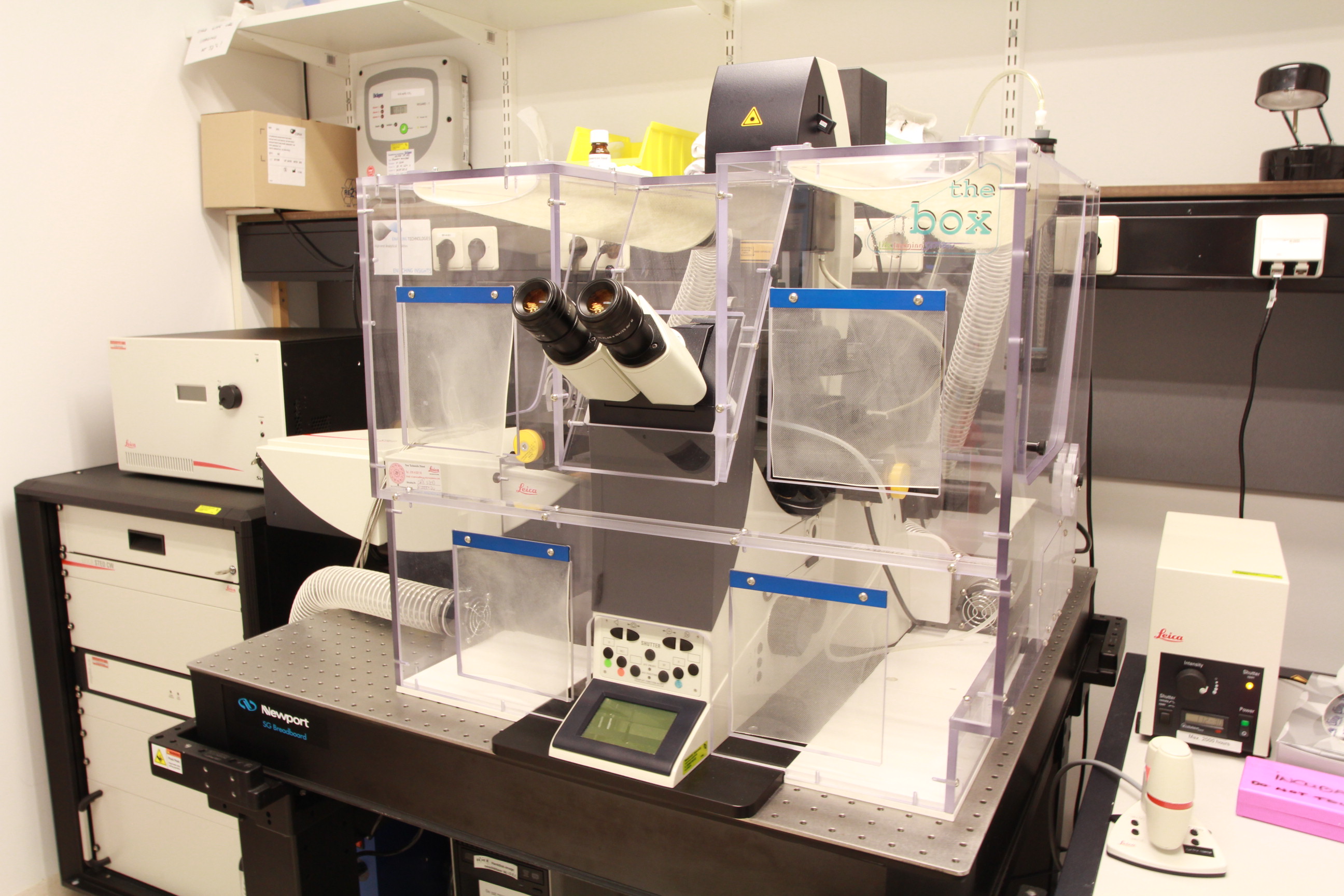
The Leica TCS SP8 STED is a fluorescence confocal laser scanning microscope with a stimulated emission depletion (STED) laser at 592nm for super-resolution fluorescence microscopy. Besides the latter depletion laser, it also contains a UV (405nm) and white-light laser (WLL2; tunable from 470 to 670nm) to excite a wide variety of fluorescent proteins and probes. For emission detection, it has two conventional PMTs as well as two hybrid HyD detectors with gating. Fast image 3D image scanning can be achieved by combining the resonance scanner and Super Z Galvo stage, whereas the CO2 and temperature controlled incubator enables live-cell imaging. The microscope runs the Leica LAS X software and acquired images can be processed directly with the SVI Huygens deconvolution software.
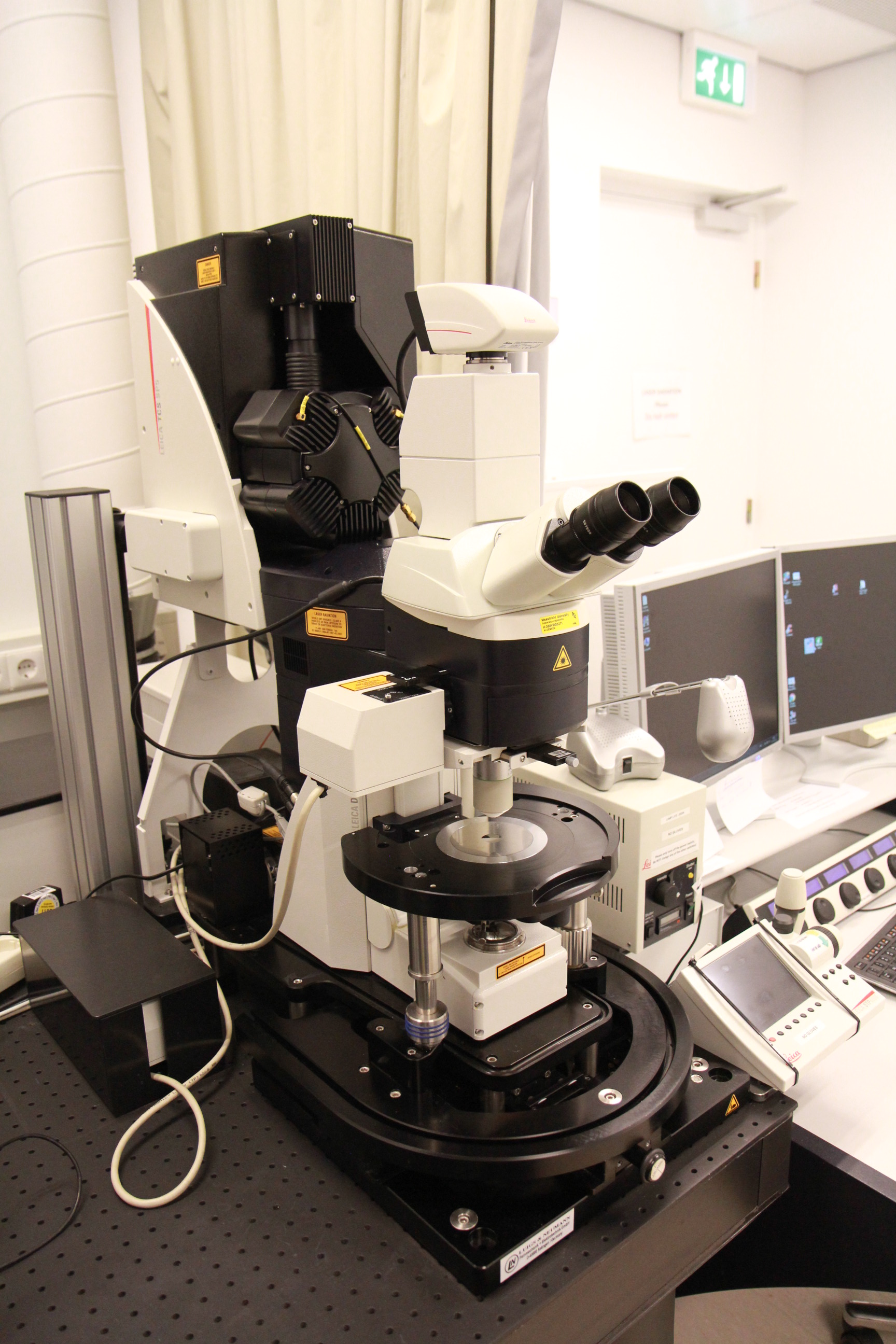
The Leica TCS SP5 MP is a fluorescent confocal laser scanning microscope with a Coherent Inc. Chameleon Ultra II Ti:sapphire laser (tunable from 680 to 1080nm) and Leica NND (non-scanned) detectors to image both deeply into tissues by two-photon excitation and image stain-free by second-harmonic generation. Forward detector? Furthermore, it contains an RGB laser for standard confocal imaging and Hamamatsu R9624 Photomultiplier tubes for detection. The Becker & Hickl Simple-Tau 830 has been installed for fluorescence life-time imaging (FLIM). For widefield microscopy, a Leica EL6000 external light source and DFC369 FX 1,4 Mpx camera can be used. The microscope stage and the room are designed and approved for animal experiments. The microscope runs the Leica LAS X software and acquired images can be processed directly with the SVI Huygens deconvolution software.
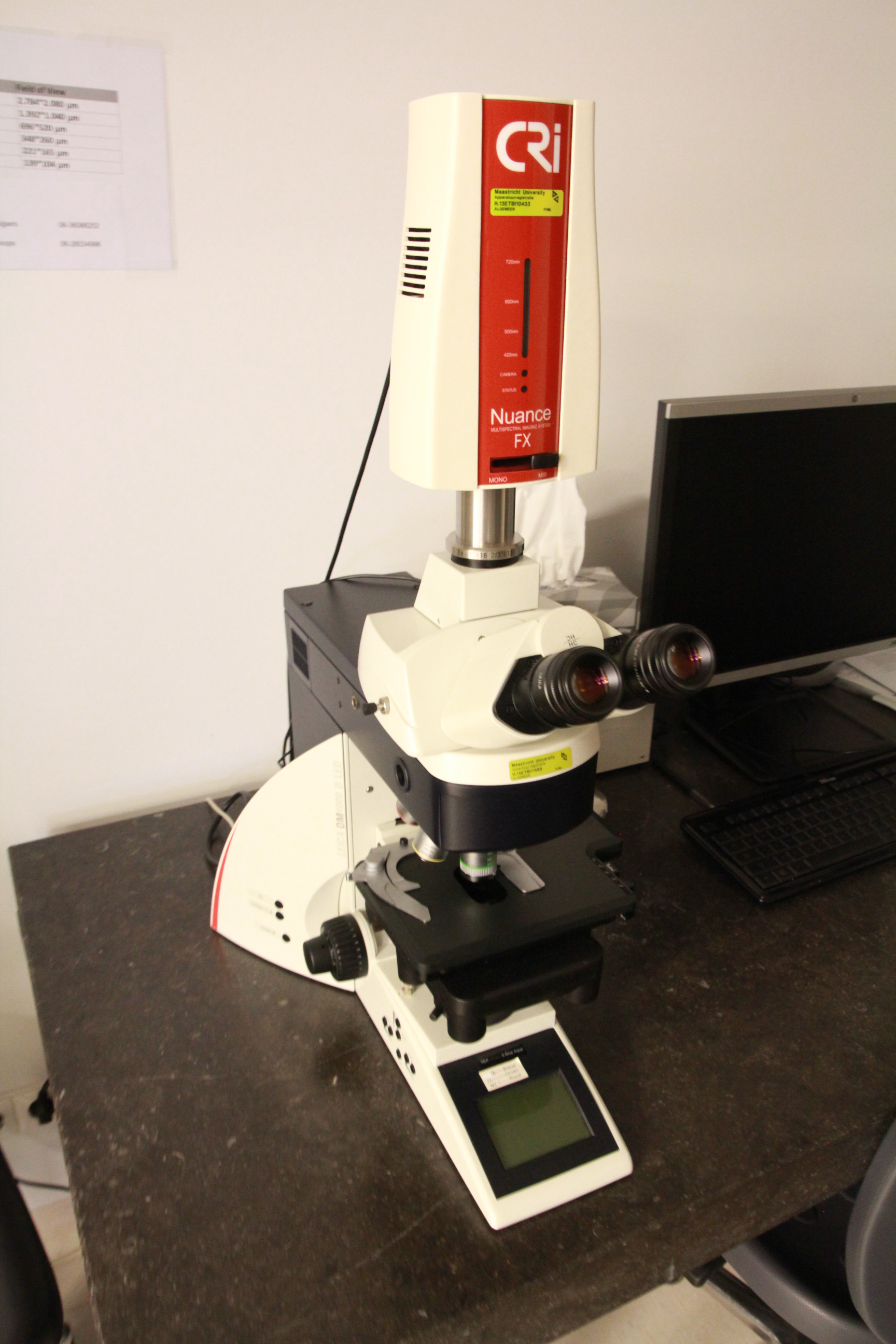
The Leica DM4000 B LED is an automated upright widefield fluorescence microscope with a manual stage and Leica EL6000 light source. It is equipped with a Perkin Elmer Nuance FX multispectral tissue imager that can be used for filter-free spectral unmixing of (auto-) fluorescence.
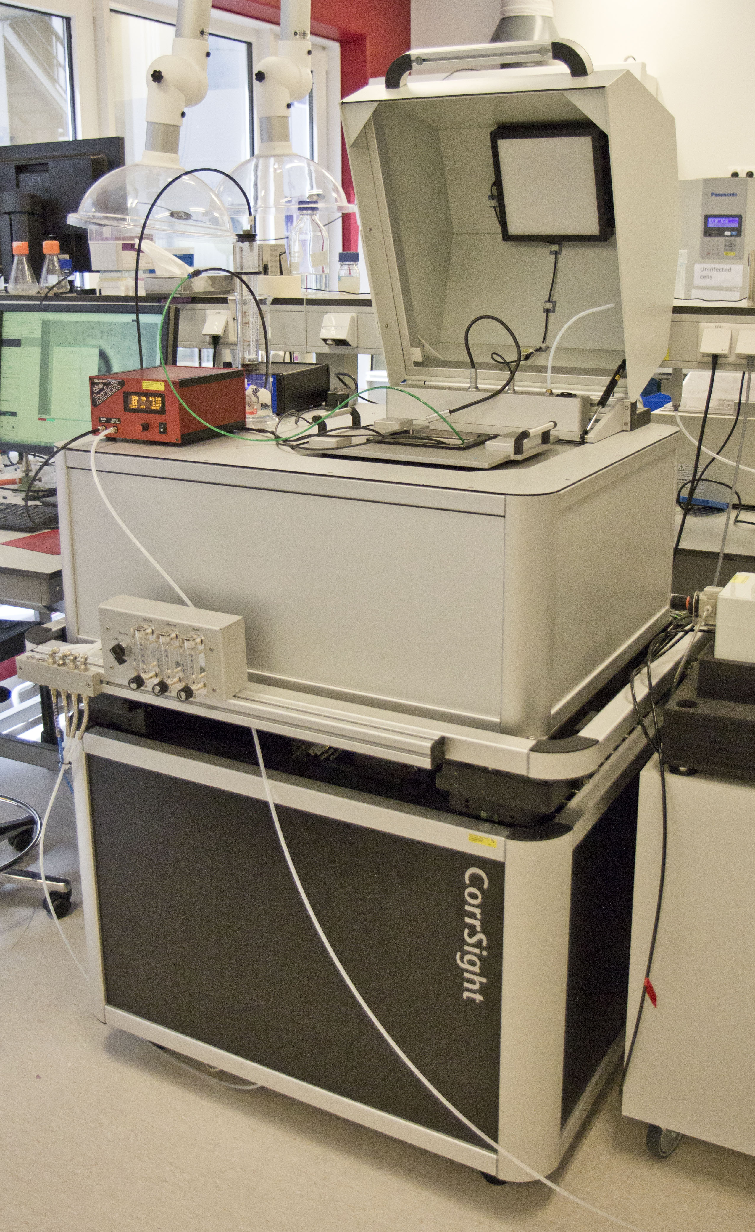
The FEI CorrSight is a (cryo-) fluorescence microscope specifically designed for correlative microscopy experiments and can be used in both widefield and spinning-disc imaging mode. Widefield imaging is achieved by a TILL Oligochrome light source, DAPI-FITC-TxRed Tripleband HC Filter and Hamamatsu 1k ORCA-R2, whereas the spinning-disc mode makes use of a Toptica iChrome MLE LFA laser module (405, 488, 561 and 640nm), an Andromeda Yokogawa spinning disk module and Hamamatsu 2k Flash 4V2plus camera. The CorrSight cryostage enables observation of samples at cryogenic temperatures, whereas the Ibidi heated chamber plate and pump enable live-cell experiments. The microscope runs the FEI MAPS software to overlay correlative fluorescence microscopy images directly onto SEM images.
The currently available objectives of all microscopes can be found here.
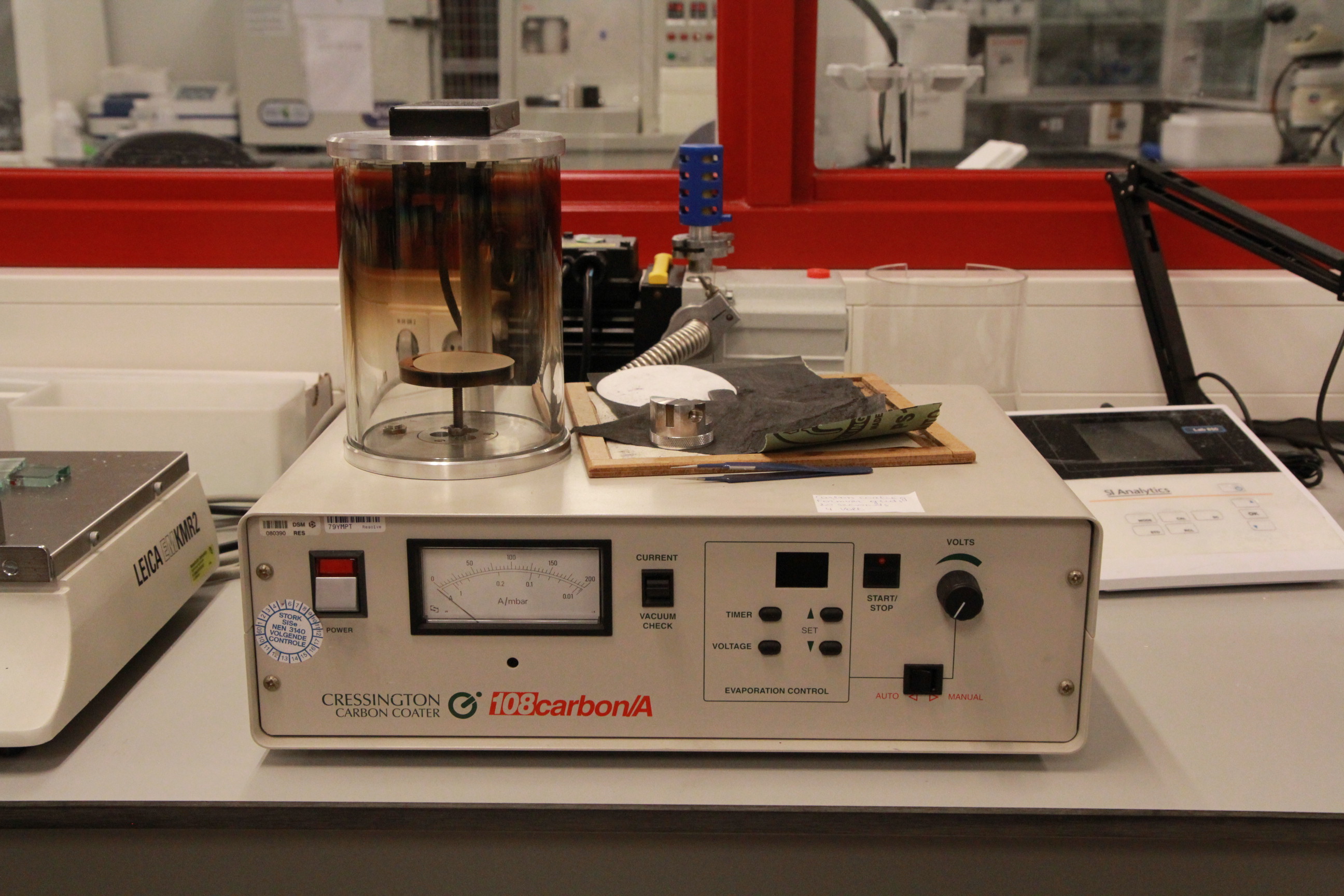
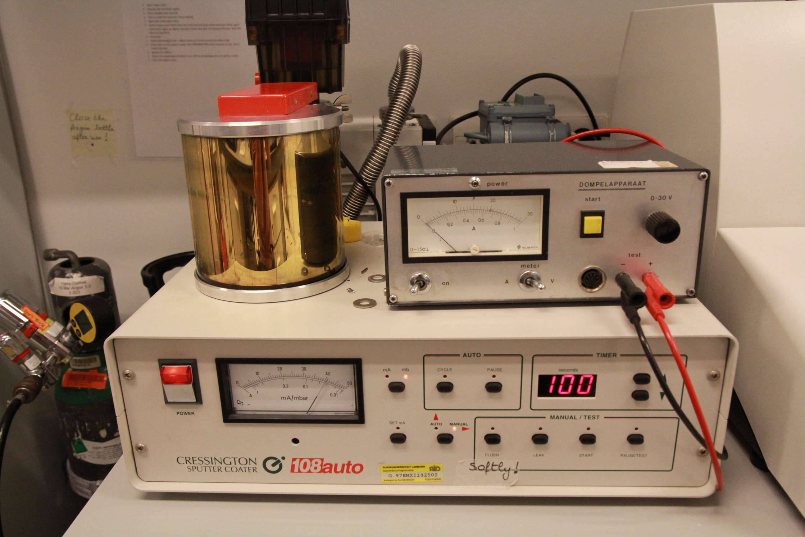
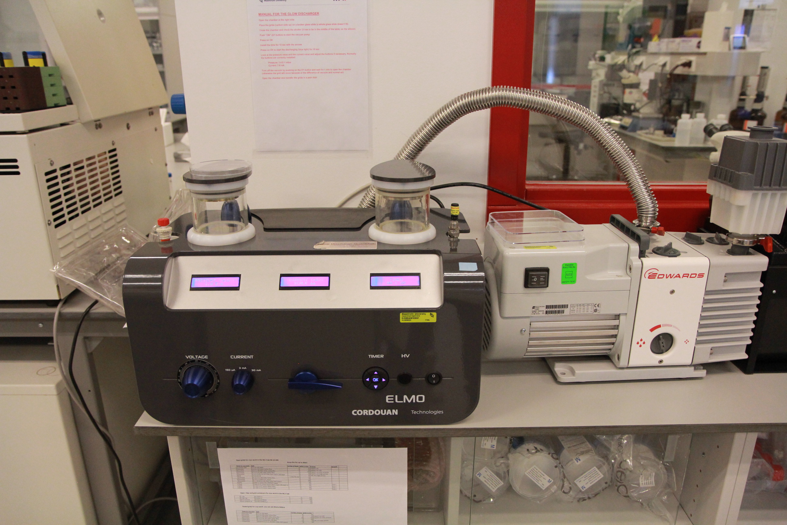
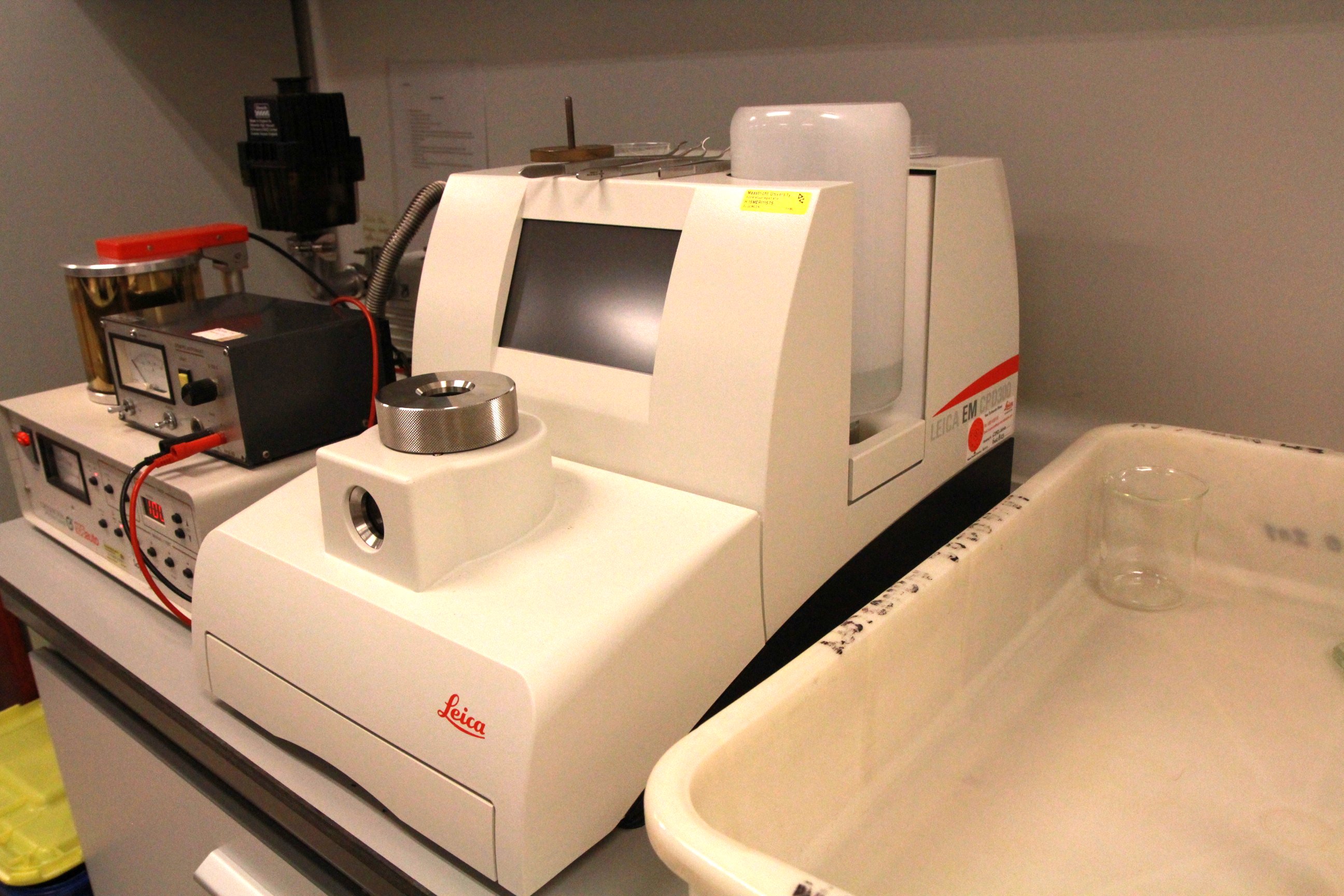
Sample preparation
Software
- FEI EM EPU and TOMO4
- EM Mesh
- FEI Amira 3D
- FEI MAPS
- inForm Cell Analysis
- Leica Las X
- SVI Huygens Professional
